CNS Imaging
Brain death
Technique
- Can be done with Tc-99m DTPA/Pertechnetate/ECD/HMPAO
- DTPA cheaper
- HMPAO or ECD do not require a flow study
Pitfalls
- Might see low-level sagittal sinus activity without obvious arterial phase possibly due to scalp vessels draining into the sinus or a small amount of intracerebral flow
- Controversial
- Regardless, no perfusion on the angiographic arterial phase have a grave prognosis if not already brain dead
- Can also see scalp perfusion which can be mistaken for intracerebral flow
- Can place elastic band around the head above orbits to diminish flow to overlying superficial scalp vessels
Positive brain death scan
- "Hot nose" sign can be caused by various things, but can be used as a secondary sign when intracerebral perfusion is absent
- Caused by increased or collateral flow through maxillary branch of external carotid due to cessation of intracranial carotid flow
- Only flow on angiographic phase should just be scalp vessels
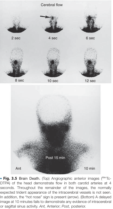
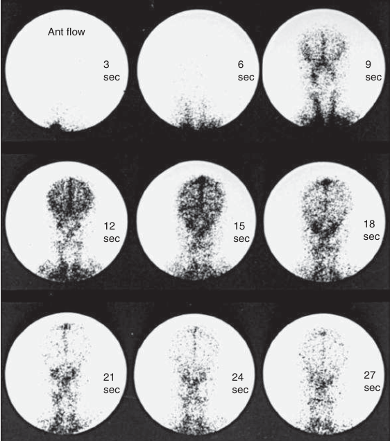
Normal anterior radionuclide angiogram:
- See trident appearance of ACAs and MCAs
- In brain death trident is absent
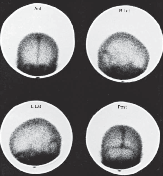
Normal planar static images
Epilepsy
- Ictal imaging more sensitive for identification of seizure foci
- Increased blood flow to this foci during seizure
- Use brain perfusion agents like Tc-99m HMPAO vs ECD
- Interictal imaging will see decreased activity
- SPECT not as sensitive so use FDG PET in these cases to look for hypometabolism
Neurodegenerative diseases
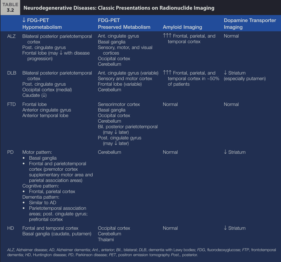
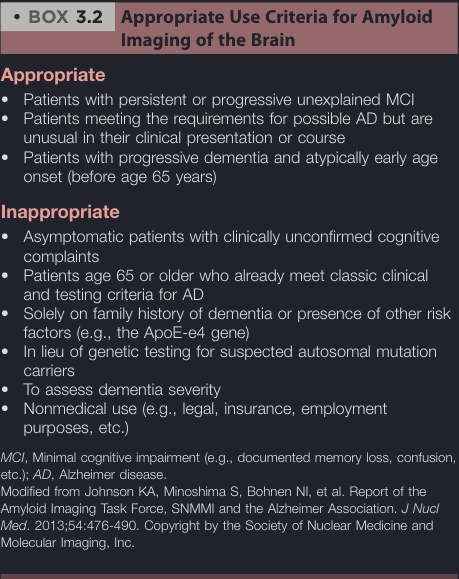
Amyloid imaging